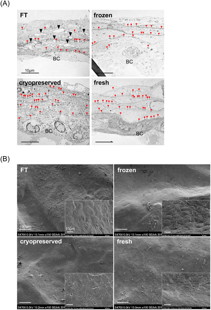
Fig 3. Electron microscopic survey of prepared cultured epidermis.
A: Transmission electron microscopy (TEM): 5–6 layers of keratinocytes, including the monolayer basal cell and desmosomal complexes (small red arrowheads) at cell-contact points, were observed in all CEs. Intercellular adhesion was rigid in fresh, cryopreserved, and frozen CEs compared with that in FT. FT: numerous small gaps were found at the intercellular space, which indicates cell membrane injury (▲). BC: basal cell. B: Scanning electron microscopy (SEM): all CEs had a similar appearance, i.e., the keratinocytes formed a smooth membrane with regularity. No crack was found in the membrane surface, even in the FT sheet. FT, freeze, and thaw.

