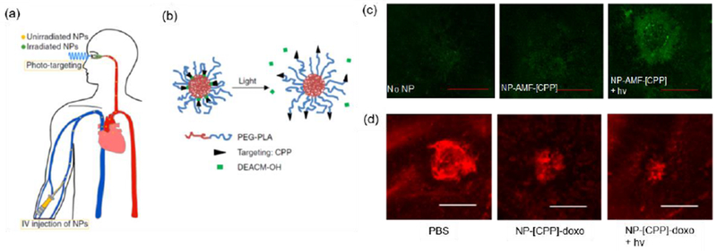
Figure 7.

Treatment of choroidal neovascularization (CNV) by photo-triggered targeting of systemically-administered nanoparticles. (a) Scheme of administration and light-triggered targeting. (b) Scheme of light-triggering of nanoparticle targeting. Irradiation removes the caging groups from the targeting motifs, which also allows them to come to the surface of the nanoparticles. (c) Targeting efficiency evaluation by confocal laser scanning microscoscopy. Accumulation of nanoparticles labeled with the fluorescent dye AMF (NP-AMF) in CNV lesions. NP-AMF-[CPP]: nanoparticles with caged cell penetrating peptide (CPP). NP-AMF-[CPP] + hv: such nanoparticles with photo-triggering. The scale bar = 100 μm. (d) Representative isolectin GS IB4-stained (red, indicating the vasculature) images of CNV lesions after treatment with doxorubicin (doxo)-containing formulations. NP-[CPP]-doxo: nanoparticles with caged CPP containing doxorubicin. Scale bar = 100 μm. Adapted from [95] with permission, copyright from Nature Publishing Group, 2019.
