
Fig. 1 |. IFN-γ-activated BMDMs secrete an antibacterial factor.
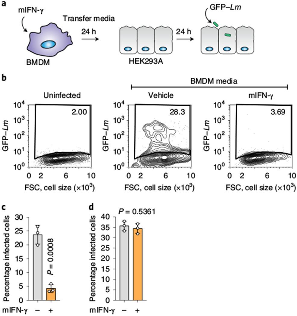
a, Schematic of the medium-transfer assay designed to investigate antibacterial products produced and secreted by mIFN-γ-stimulated BMDMs. b, HEK293A cells cultured in conditioned medium from mIFN-γ-stimulated BMDMs were infected with GFP-expressing L. monocytogenes (multiplicity of infection (m.o.i.) = 2; 22 h) as indicated. The flow cytometry plots show the percentage of GFP-positive HEK293A cells. FSC, forward scatter. c, Quantification of the assay described in b (BMDM-conditioned medium). d, HEK293A cells were not affected by residual mIFN-γ present in BMDM-conditioned media. HEK293A cells were treated with 500 U ml−1 mIFN-γ for 24 h and infected with GFP-expressing L. monocytogenes the next day (m.o.i. = 2; 22 h). Infection was quantified by flow cytometry as in c. c,d, The bars represent the mean values. The error bars show the s.d. from three independent experiments and statistical significance was determined using a Student’s unpaired t-test (two-tailed). Lm, L. monocytogenes.