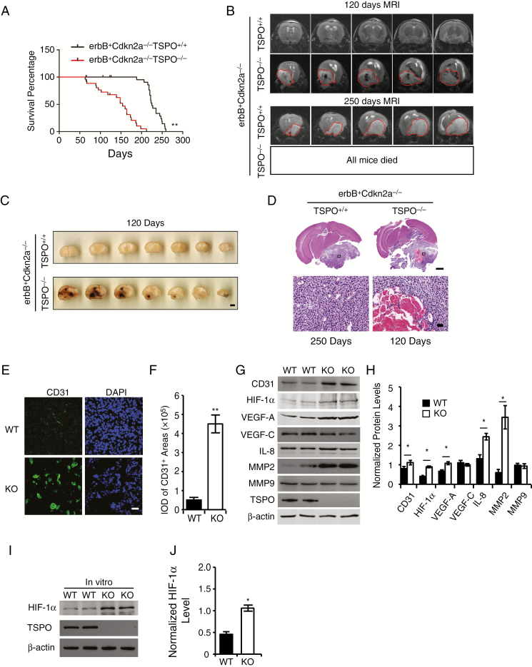
Fig. 2.
TSPO deletion promotes glioma pathogenesis and malignancy in a spontaneous primary glioma mouse model by increasing hypoxia-induced angiogenesis. (A) Survival curves for S100b-v-ErbB+Cdkn2a-/-TSPO-/- mice (n = 27) and S100b-v-ErbB+Cdkn2a-/-TSPO+/+ littermates (n = 26). (B) The upper panel shows representative MRIs of the brains of S100b-v-ErbB+Cdkn2a-/-TSPO-/- mice and S100b-v-ErbB+Cdkn2a-/-TSPO+/+ littermates at the age of 120 days. The circled regions indicate the gliomas, which appear dark, suggesting extensive hemorrhaging in the gliomas. The lower panel shows a representative brain MRI of an S100b-v-ErbB+Cdkn2a-/-TSPO+/+ mouse at the age of 250 days. The circled regions indicate the gliomas, which appear relatively white, suggesting little hemorrhaging. No brain MRIs were obtained from S100b-v-ErbB+Cdkn2a-/-TSPO-/- mice because all of these mice died before 210 days. (C) Representative images of a series of slices from S100b-v-ErbB+Cdkn2a-/-TSPO-/- mice and S100b-v-ErbB+Cdkn2a-/-TSPO+/+ littermates at the age of 120 days. Scale bar, 2 mm. (D) Representative images of HE-stained brain slices from S100b-v-ErbB+Cdkn2a-/-TSPO-/- mice (120 days) and S100b-v-ErbB+Cdkn2a-/-TSPO+/+ littermates (250 days). Scale bar, 1 mm for upper images and 100 μm for lower images. (E) Representative images of CD31 immunofluorescence staining of glioma tissues. Scale bar, 20 μm. (F) Quantification of CD31 levels in glioma tissues. (G) Western blot analysis of the levels of the angiogenesis-associated proteins in glioma tissues. (H) Quantification of the relative levels of angiogenesis-associated proteins shown in panel G compared with β-actin. (I) Western blot analysis of HIF-1α levels in TSPO-KO cells compared with WT cells. (J) Normalized HIF-1α levels in panel I. Three independent experiments were performed. The data are presented as means ± SEM. *P < 0.05 and **P < 0.01 as determined using Student’s t-test (F, H, and J) or the Gehan-Breslow-Wilcoxon test (A).

