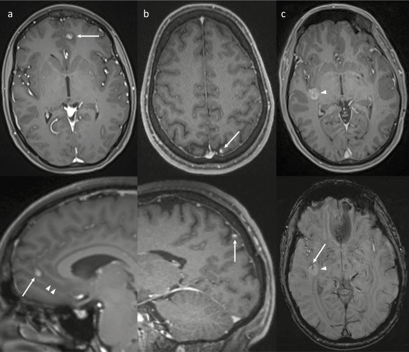
Fig. 3.
Examples of using other planes and sequences (bottom images) to improve characterization of IMM identified on axial post-contrast imaging (top images). (a) The sagittal reconstruction (bottom) better demonstrates an elongated morphology of the IMM (arrows) being oriented along a sulcus (arrowheads). (b) The sagittal plane (bottom) helps confirm that the IM metastasis (arrows) is oriented along the corticomeningeal interface. (c) Susceptibility weighted imaging (bottom) shows a vessel within the central sulcus (arrow) passing into the middle of the IMM (arrowhead), showing that the IM metastasis surrounds the central sulcus. This relationship is not as well appreciated on the postcontrast image (top).

