
Figure 2: Potential mechanisms resulting in lower tissue NAD+ concentrations with age.
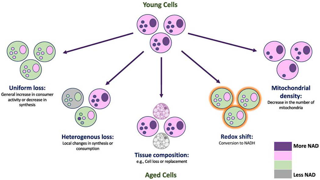
Schematic illustrating distinct scenarios that could result in lower measured NAD+ in extracts of tissues from aged animals. Uniform loss involves all cells experiencing a similar NAD+ deficit. Heterogenous loss suggests local defects resulting in impaired synthesis or excess consumption that could affect a subset of cells disproportionately. Tissue composition may also change with age, resulting in decreased cellularity or the appearance of cells with less NAD+ (e.g., adipocytes). A shift in the redox balance could lower NAD+ without any change in the total (NAD+ + NADH) pool. A decrease in the number of mitochondria (or other NAD+-rich organelles) could decrease the whole-tissue NAD+ concentration and apparent redox state (i.e., whole tissue NAD+:NADH ratio) without actually changing NAD+ concentration or redox state in any given compartment.