
Figure 3. HDAC10 regulates tumor pluripotency.
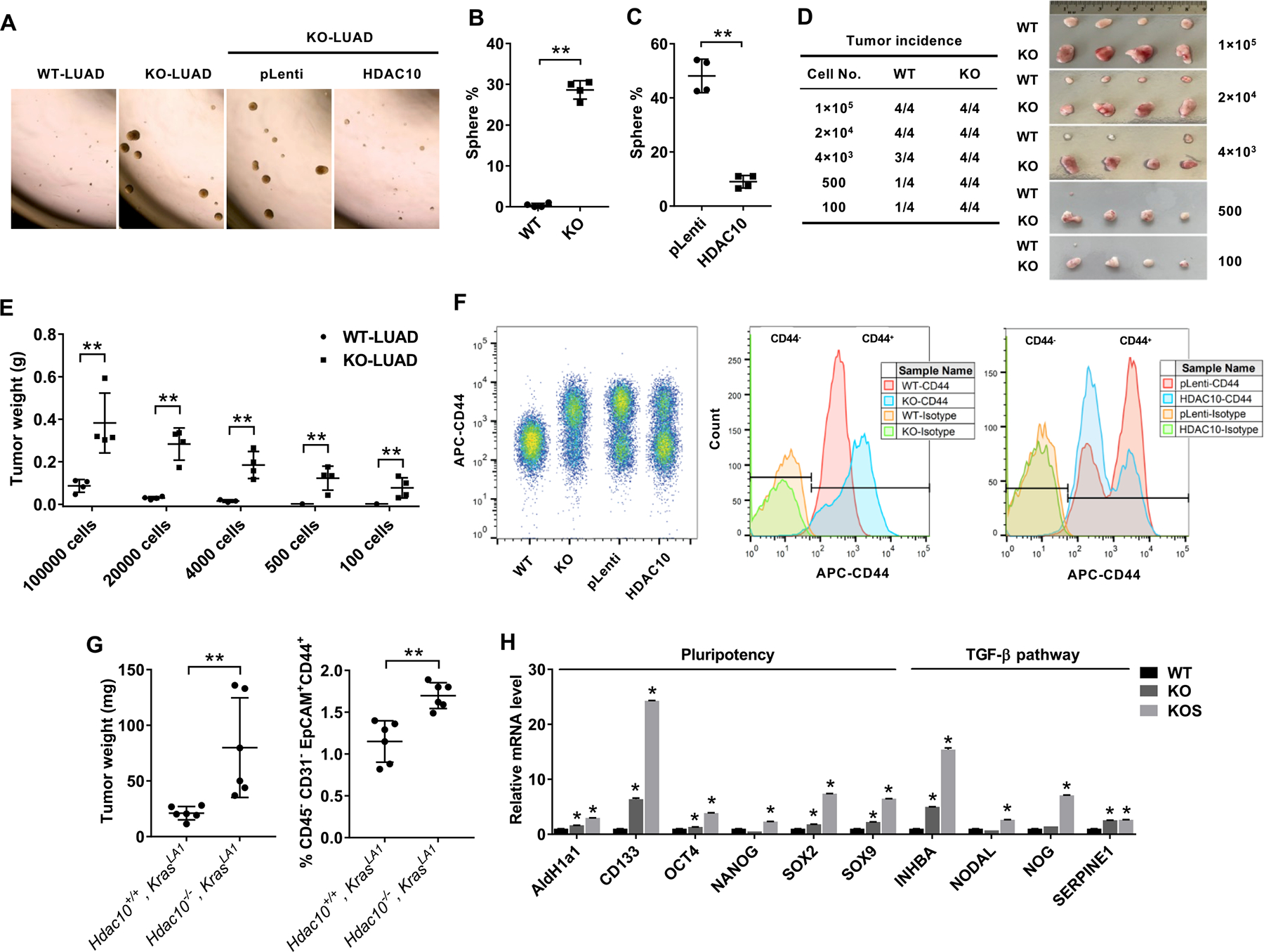
(A) Tumor sphere cultures of mouse LUAD cells 1 week after plating in a 24-well ultra-low attachment plate. (B) Quantification of the percentage of tumor spheres from Hdac10 WT and KO LUAD cells. (C) Quantification of tumor spheres from Hdac10 KO LUAD cells transduced with lentiviral control (pLenti) or Hdac10 expression vectors (HDAC10). (D) Tumor incidence of the in vivo limiting dilution assay. Representative image of tumors (right panel) and tumor incidence (left panel) at 3 weeks were indicated. (E) The tumors of the indicated groups from (D) were weighed. (F) Flow cytometric analysis of CD44 expression in LUAD cells. CD44 expression was increased in Hdac10 KO LUAD cells, whereas overexpression of HDAC10 in Hdac10 KO LUAD cells caused a decrease of CD44high population. (G) Flow cytometry showed a markedly increased CD45−CD31−EpCAM+CD44+ population in lung tumors from Hdac10−/−, KrasLA1mice at 30 weeks (right panel). (Left panel) The weights of total lung tumors from mice were indicated. (H) Results of real-time RT-PCR analysis of the expression of pluripotency transcription factors and TGF-β pathway in Hdac10 WT and KO LUAD, as well as KO LUAD spheres (KOS). *, P < 0.05 or **, P < 0.01.