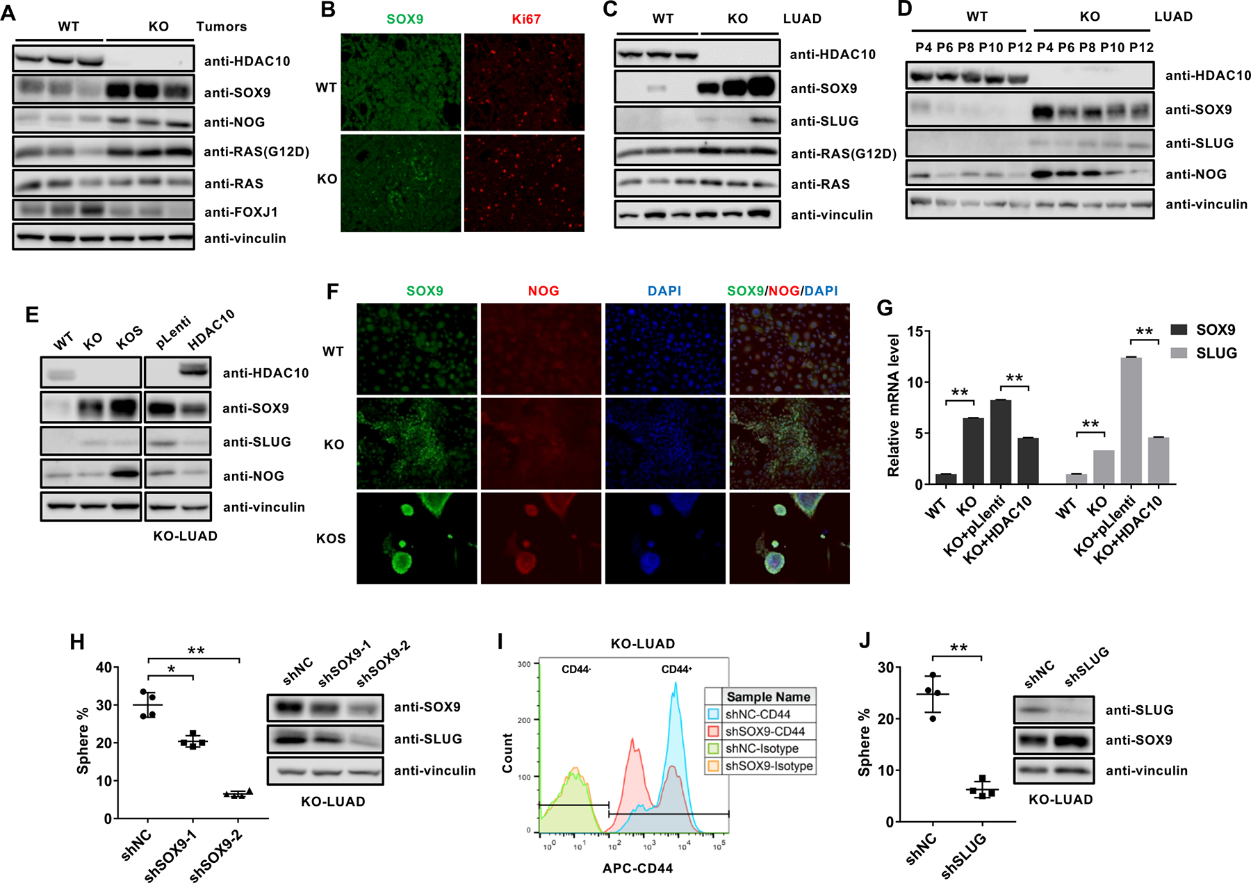
Figure 4. SOX9 is upregulated in Hdac10-deleted tumor cells.

(A) Western analysis of the indicated proteins in Hdac10 WT and KO tumor tissues from mice at 30 weeks. (B) Immunostaining of SOX9 (green) and Ki67 (red) from mice at 30 weeks. (C) Western analysis of the indicated proteins in Hdac10 WT and KO LUAD cells isolated from different mice. (D) Western blot analysis of SOX9, SLUG and NOG expression in Hdac10 WT and KO LUAD cells with indicated passage number. (E) Western blot analysis results for the indicated proteins in Hdac10 WT, KO and KO spheres (KOS) LUAD cells (left panel), or Hdac10 KO LUAD cells transduced with the control vector or vector encoding HDAC10 (right panel). (F) Immunostaining of SOX9 (green), NOG (red) and DAPI (blue) in LUAD cells. (G) Results of real-time RT-PCR analysis of the expression of SOX9 and SLUG. (H) The effect of Sox9 depletion on the growth of tumor spheres in Hdac10 KO LUAD cells. Quantification of tumor spheres (left panel) and Western analysis (right panel) of SOX9 and SLUG in Hdac10 KO LUAD cells transduced with control (shNC) or SOX9 shRNA lentiviral vector (shSOX9–1 and shSOX9–2). (I) Flow cytometric analysis of the effect of SOX9 depletion on CD44 expression in Hdac10 KO LUAD cells. (J) The effect of SLUG depletion on the growth of tumor spheres in Hdac10 KO LUAD cells. *, P < 0.05 or **, P < 0.01.
