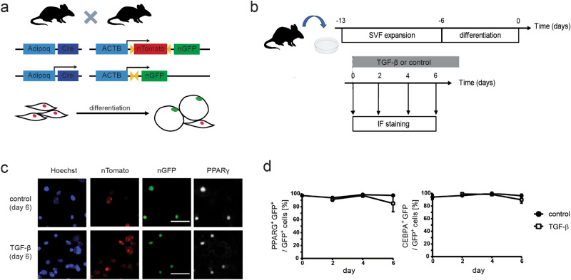
Figure 1.
TGF-β stimulation does not induce the loss of adipocyte marker expression under standard culture conditions in primary mouse adipocytes differentiated ex vivo. (a) Schematic of the transgenic mouse model used. (b) Experiment outline to test the effect of TGF-β on primary adipocytes using immunofluorescent detection of GFP and adipocyte markers PPARγ and C/EBPα. Primary SVF cells from Adipoq:Cre nT/nG mice were expanded and differentiated into adipocytes in vitro. TGF-β was added to the culture media at the end of differentiation (day 0) and cells were analyzed at days 0, 2, 4 and 6 using immunofluorescent staining. (c) Representative fluorescent images of staining against PPARγ at day 6 after adding stimulus. GFP expression is colocalized with PPARγ expression in the nuclei of both control and TGF-β-treated cells. Scale bar: 50 µm. (d) Percentage of GFP-positive cells expressing adipocyte markers PPARγ and C/EBPα. Two-tailed Student t tests with Benjamini–Hochberg correction; FDR = 0.01; n = 3–8 technical replicates, all time points p > 0.05.

