
Figure 2. Neutralization of SARS-CoV-2 pseudovirus by plasma.
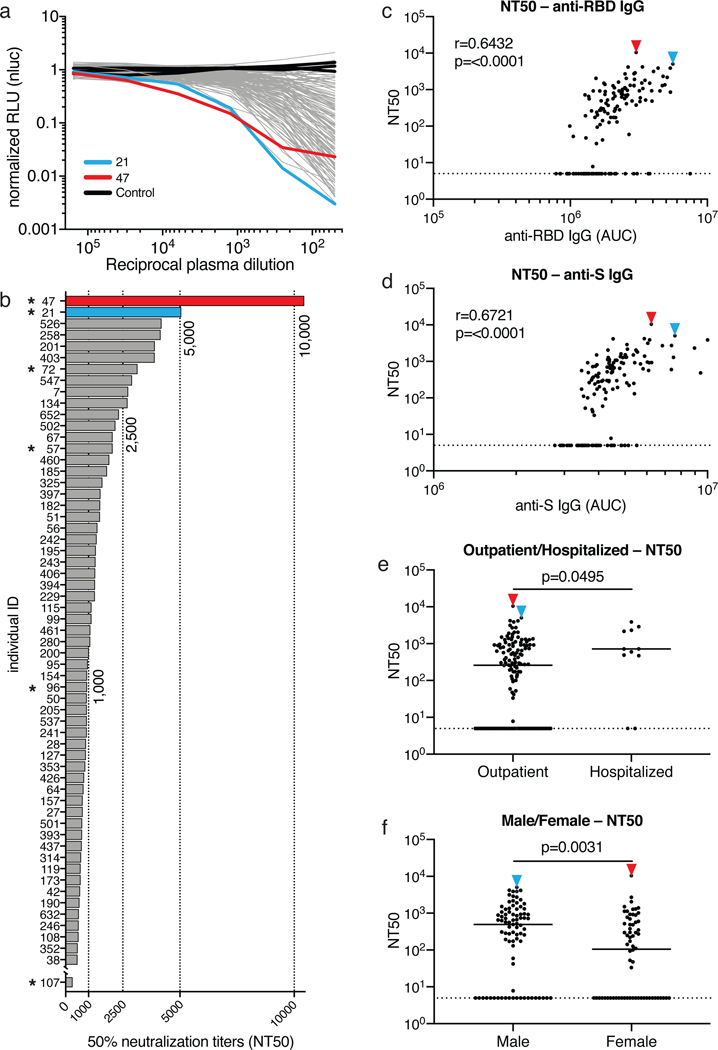
a, Graph shows normalized relative luminescence values (RLU, Y axis) in cell lysates of 293TACE2 cells 48 hours after infection with nanoluc-expressing SARS-CoV-2 pseudovirus in the presence of increasing concentrations of plasma (X axis) derived from 149 participants (grey, except individuals 21 and 47 in blue and red lines, bars and arrowheads, respectively) and 3 negative controls (black lines). Mean of duplicates; representative of two independent experiments. b, Ranked average half-maximal inhibitory plasma neutralizing titer (NT50) for the 59 of 149 individuals with NT50s >500 and individual 107. Asterisks indicate donors from which antibody sequences were derived. c, Normalized AUC for anti-RBD IgG ELISA (X axis) plotted against NT50 (Y axis); r=0.6432, p=<0.0001. d, Normalized AUC for anti-S IgG ELISA (X axis) plotted against NT50 (Y axis); r=0.6721, p=<0.0001. The r and p values for the correlations in c and d were determined by two-tailed Spearman’s. e, NT50 for outpatients (n=138) and hospitalized (n=11) individuals; p=0.0495. f, NT50 for males (n=83) and females (n=66) in the cohort; p=0.0031. Statistical significance in e and f was determined using two-tailed Mann-Whitney U test and horizontal bars indicate median values. Dotted lines in c to f (NT50=5) represents lower limit of detection (LLOD). Samples with neutralizing titers below 1:50 were plotted at LLOD.