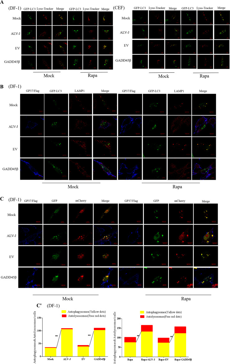
Fig. 3. Autophagosomes are unable to fuse with lysosomes following ALV-J infection or the overexpression of GADD45β.
a DF-1 cells or CEF cells were marked with Lyso Tracker Red (50 nM) for 2 h and GFP-LC3B-labeled autophagosomes (green) were visualized using confocal microscopy to colocalize red-stained acidified vesicles (red). b Cells were stained with anti-LAMP1 antibody (red) and anti-gp37 antibody (blue), and visualized using confocal microscopy to visualize the fusion between the autophagosomes and lysosomes. c Cells were infected with the mCherry-GFP-LC3B recombinant adenovirus to analyze the smoothness of autophagosome formation. Scale bar, 10 μm. The chart (c′) shows the quantification of GFP-mCherry-LC3 tandem reporter in (c).

