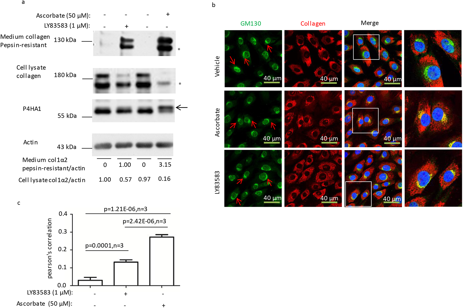
Figure 3. LY83583 stimulated collagen secretion is independent of P4HA1 N259 glycosylation.

(a) Ascorbate but not LY83583 treatment for 6 hrs induced N259 glycosylation on P4HA1 (panel 3, indicated by an arrow) and more pepsin-resistant Type I collagen in the culture medium (panel 1). (b and c) Ascorbate treatment induced significantly more type I collagen (red) entering Golgi apparatus (green stained by GM130, indicated by red arrows). The higher resolution parts in the panels were indicated by the squares. Statistical analysis was performed from three different immunofluorescence staining experiments. Co-localization index (Pearson’s correlation) was calculated based on NIS-Elements AR software. Collagen in the culture medium or lysate to actin was quantified by IMAGE J software. * indicated pepsin-resistant col 1 α2. P values were indicated in each panel if available.
