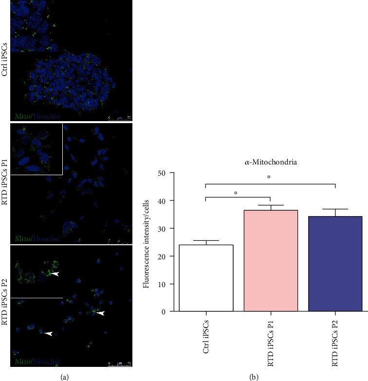
Figure 5.

Confocal analysis of MTCO2 (Mito) in Ctrl and RTD cells: (a) immunofluorescence images of the mitochondrial marker (in green) demonstrating the higher abundance of organelles in patients' cells, as compared to controls. White arrowheads, immunoreactive aggregates; (b) fluorescence intensity levels reveal a significant increase (∗p ≤ 0.05) of α-mitochondria in RTD cells vs. Ctrl iPSCs. Nuclei are stained with Hoechst (in blue).
