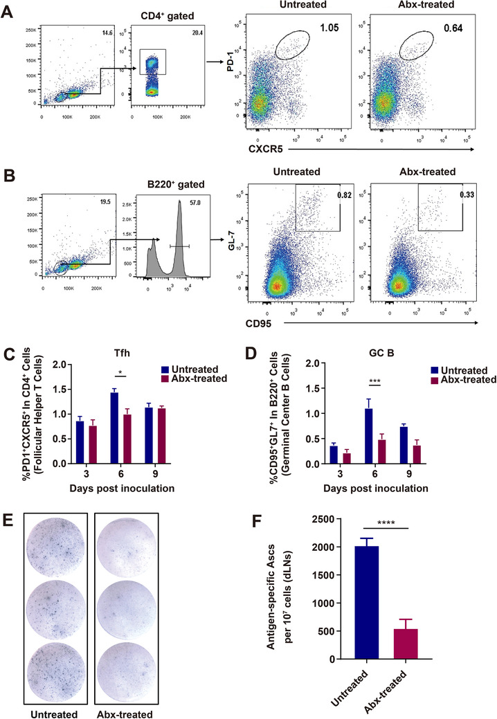
FIGURE 2.

Recruitment of Tfh, GC B cells, and ASCs in Abx‐treated and untreated mice immunized with rabies vaccine. A‐D, Tfh and GC B cells in the draining LNs (dLNs) post rabies vaccination. Abx‐treated and untreated mice were inoculated i.m. with 107 FFU iLBNSE. dLNs were harvested, and single‐cell suspensions of the dLNs were stained with Tfh and GC B cell markers, and analyzed by flow cytometry (Untreated mice, n = 5, Abx‐treated mice, n = 5). A, Representative gating strategy for identification of Tfh cells. B, From the dLNs, the percentage of PD1+ CXCR5+ Tfh cells in CD4+ B cells. Error bars in the graphs represent standard error (* P < .05; Student's t‐test). C, Representative gating strategy for identifying GC B cells. D, From the dLNs, percentage of GL7+ CD95+ GC B cells in B220+ B cells. Error bars in the graphs represent standard error (*** P < .001; Student's t‐test). E,F, Quantification of ASCs in the draining LNs of Abx‐treated and untreated mice post immunization. Single‐cell suspensions of the dLNs were added into ELISpot plates coated with purified RABV, and subsequently incubated with Biotin‐mIgG Ab and Streptavidin‐Alkaline Phosphatase, signal was detected with BCIP/NBT‐plus (Untreated mice, n = 5, Abx‐treated mice, n = 5). E, Representative ELISpot images of ASCs from the draining LNs. F, Graph of ELISpot results. Error bars represent standard error ( ****P < .0001; Student's t‐test)
