
Figure 3.
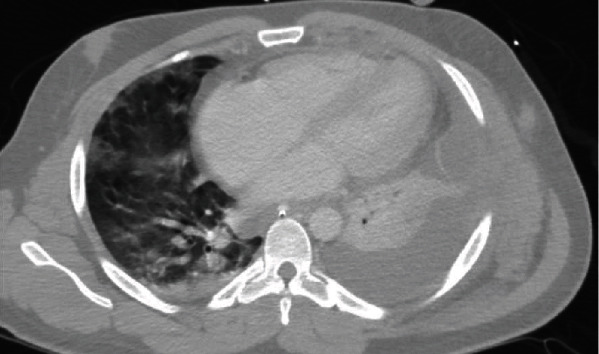
CT chest shows left-sided pleural effusion with loculations and atelectasis. Nodular groundglass opacities are present in the right upper and middle lobes. There is a demonstration of a small pericardial effusion.
Official websites use .gov
A
.gov website belongs to an official
government organization in the United States.
Secure .gov websites use HTTPS
A lock (
) or https:// means you've safely
connected to the .gov website. Share sensitive
information only on official, secure websites.
CT chest shows left-sided pleural effusion with loculations and atelectasis. Nodular groundglass opacities are present in the right upper and middle lobes. There is a demonstration of a small pericardial effusion.