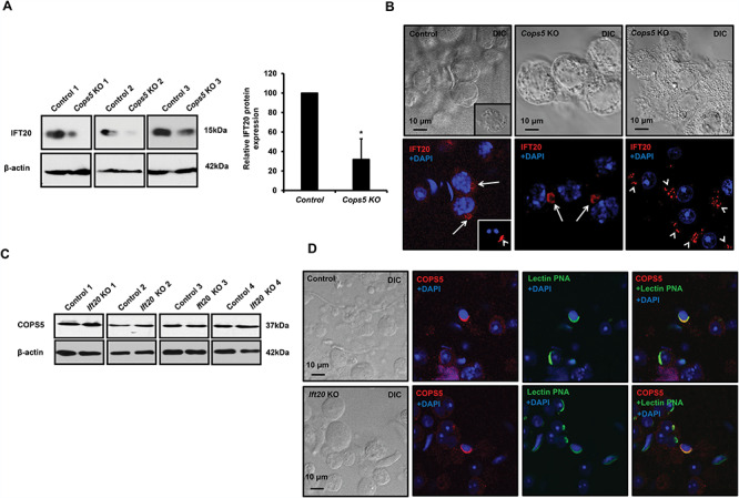
Figure 7.

COPS5 determines IFT20 expression level and localization in male germ cells, but COPS5 localization and expression were not changed in the Ift20 knockout mice. A. Analysis of testicular IFT20 expression in control and Cops5 knockout mice by Western blot. The expression level of IFT20 was reduced remarkably in the knockout mice (n = 3). B. IFT20 localization in germ cells of the control and Cops5 knockout mice by immunofluorescence staining (red). IFT20 was localized in the Golgi body of a spermatocyte (arrows) and acrosome of a round spermatid (arrow heads). In the Cops5 knockout mouse, IFT20 was still present in the Golgi body of the spermatocyte but was no longer seen in the spermatid acrosome. However, the protein was present as small vesicles near one side of the nucleus. C. Expression levels of COPS5 in control and Ift20 conditional knockout mice. Compared with control mice, there was no change in the COPS5 expression level; D. COPS5 was still present in the acrosome of spermatids from the Ift20 knockout mice. The testicular cells were double stained with COPS5 antibody (red) and the acrosome marker lectin PNA (green). Like in the control mouse, COPS5 was still present in the acrosome. Unlike the Cops5 knockout mice, acrosome staining was not affected in the Ift20 knockout mice.
