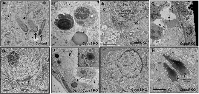
Figure 8.

Ultra-structural changes in the seminiferous tubules of Cops5 knockout mice. A. Normally developed elongating spermatids (arrows) in a control mouse. B. Normally developed round spermatid in a control mouse, showing a well-developed acrosome (arrow) and a prominent Golgi apparatus (G). C. Representative apoptotic germ cells in the seminiferous epithelium of the Cops5 KO. D. Apoptotic cells are being engulfed by Sertoli cells. The upper arrow is pointing to a Sertoli cell nucleus. The black debris is the left-over material after digestion (lower arrow). E. Step 4 round spermatid showing failure of acrosome formation in the flattened area under the Golgi apparatus (G), where the acroplaxome is also missing. F. A round spermatid in the Cops5 KO that has a thin acrosome but formed an abnormal ectoplasmic specialization (ES) with the Sertoli cell. G. The arrows point to vacuoles, presumably the space left remaining after apoptotic cells have been phagocyted by Sertoli cells. H. Two elongated spermatids that formed what appear to be normal acrosomes, but show abnormal nuclear structures.
