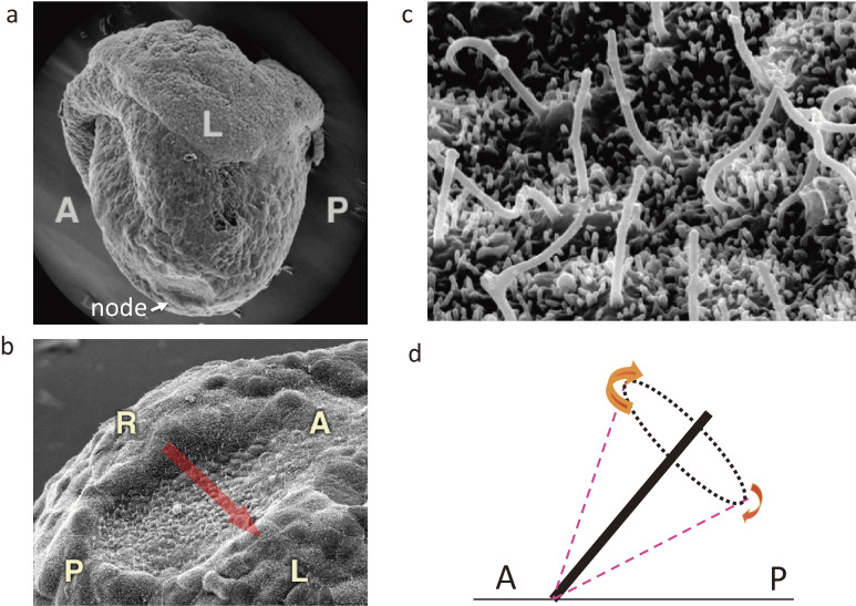
Figure 3.
Cilia at the node of a mouse embryo as revealed by scanning electron microscopy. a. Lateral view of a mouse embryo at E7.5. The arrow indicates the location of the node. b. Ventral view of the mouse node. The arrow indicates the direction of fluid flow at the node. c. Higher magnification of the node showing the presence of motile cilia. d. Posterior tilt of motile cilia. Clockwise rotation of posteriorly tilted cilia generates fluid flow (leftward) most efficiently when the cilia are farthest from the surface. A, anterior; L, left; P, posterior; R, right. Modified with permission from Shiratori and Hamada.155)

