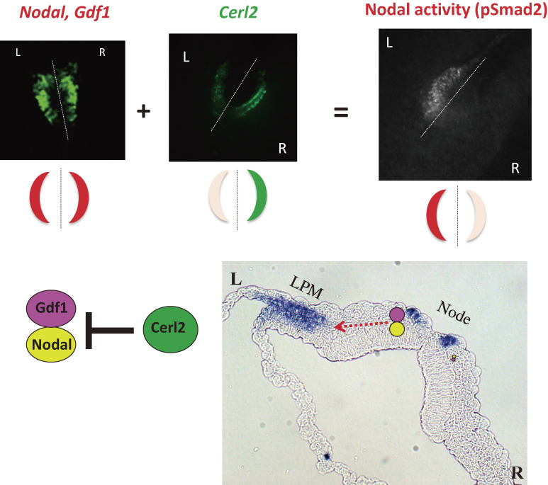
Figure 6.
Generation of molecular asymmetries at the node. Whereas Nodal mRNA and Gdf1 mRNA are present at similar levels on both sides of the node of a mouse embryo, Cerl2 mRNA shows an asymmetric (R > L) distribution (top panels). Nodal and Gdf1 form a heterodimer that constitutes an active form of Nodal. Given that Cerl2 is an inhibitor of Nodal (bottom left panel), the level of Nodal activity, which is reflected by the abundance of phosphorylated Smad2/3 (pSmad2), shows a R ≪ L pattern (top panels). The Nodal-Gdf1 heterodimer produced by and secreted from perinodal crown cells is thought to be transported to the LPM on the left side via an intraembryonic route (red dotted arrow in the bottom right panel). On reaching the LPM, the Nodal-Gdf1 heterodimer is thought to activate expression of Nodal (indicated by purple staining in the bottom right panel), which is responsive to Nodal signaling. Modified with permission from Shiratori and Hamada,155) Yoshiba and Hamada,156) and Shiratori and Hamada.157)

