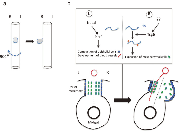
Figure 12.
Rotation and looping of the developing gut. a. Clockwise rotation of the foregut results in translocation of the liver primordium (gray) to the right side of the body cavity. b. Cellular changes that initiate L-R asymmetry in the midgut tube of the chick embryo.120,123) Asymmetries arise in the dorsal mesentery between Hamburger–Hamilton (HH) stages 20 and 22. Left side-specific Nodal-Pitx2 expression drives compaction of mesenchymal cells (green rectangles) within the left dorsal mesentery and promotes retention of a columnar morphology of epithelial cells (blue rectangles) on the left side of the dorsal mesentery. In the right dorsal mesentery, hyaluronan (HA) undergoes modification by Tsg6, which catalyzes the covalent attachment of a heavy chain (orange) of inter-α-trypsin inhibitor. Signals that induce Tsg6 expression in the right half of the dorsal mesentery remain unknown. The modified form of hyaluronan is more stable and accumulates in the right dorsal mesentery, resulting in expansion of mesenchymal cells and exclusion of blood vessels (red line) in the right dorsal mesentery. Together, these cellular asymmetries drive leftward tilting of the gut tube. Left (L) and right (R) sides of the dorsal mesentery are indicated together with the midline (broken line). Modified with permission from Hamada.158)

