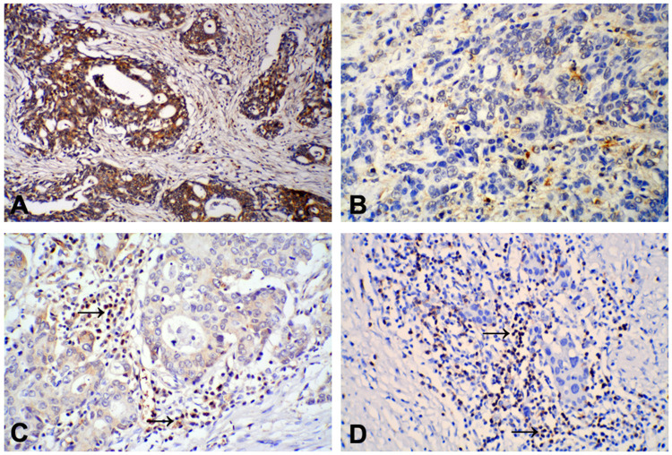
Figure 1.
Positive staining of 15-PGDH protein was mainly found in the cytoplasm of gastric cancer (GC) tumor cells: (A) High/moderately differentiated GC, positive staining; (B) Poorly differentiated GC, negative staining. IHC staining of FOXP3 expression was mainly observed in the cytoplasm of lymphocytes in gastric cancer tissues: FOXP3 expression in high/moderately differentiated GC was lower (C) than in poorly differentiated GC (D) tissues. (×200).

