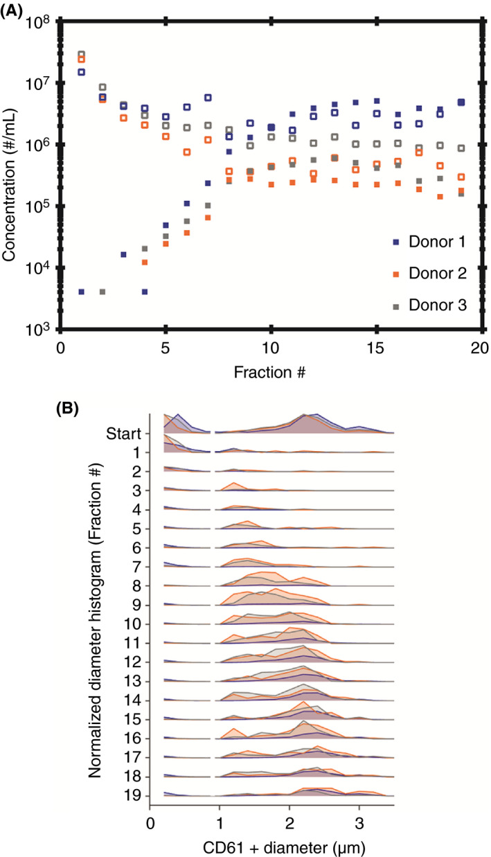
Figure 4.

Platelet and platelet‐derived extracellular vesicles (EV) concentration and diameter measured in rate zonal centrifugation fractions (RZC) for 3 donors. A, The highest concentration of platelets (closed symbols) are present in fractions 8‐17. The highest concentration of platelet‐derived EVs (open symbols) are present in fractions 1‐7. B, The platelet and platelet‐derived EV diameter in RZC fractions for donors 1‐3. The platelet diameter range (>1 µm) was obtained using Rosetta calibration. The platelet diameter range shifts to the right while moving through the gradient, which confirms that the separation of platelets from platelet‐derived EVs (200 nm to 1 µm) by RZC is based on diameter
