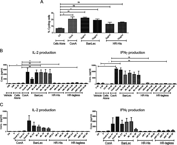
Figure 6.
Nonmitogenic activity and the absence of cytokine-inducing activity of HR. A, flow cytometry analysis of cell proliferation data of splenocytes treated with ConA, BanLec, and HR at 5 and 10 µg/ml at 36-h time points. FACS data show the amount of propidium iodide staining representing cells in different phases of the cell cycle. B, cytokine profile of the time-dependent (36- and 48-h) protein stimulation experiment by ConA, BanLec, and HR (tagged and tagless) at 10 µg/ml concentration on mouse splenocytes. Values shown are the mean from triplicate samples. C, cytokine profile of concentration-dependent stimulation (2.5–20 µg/ml) of mouse splenocytes by ConA, BanLec, and HR tagged and tagless after 48-h incubation. Values shown are the mean from triplicate samples.

