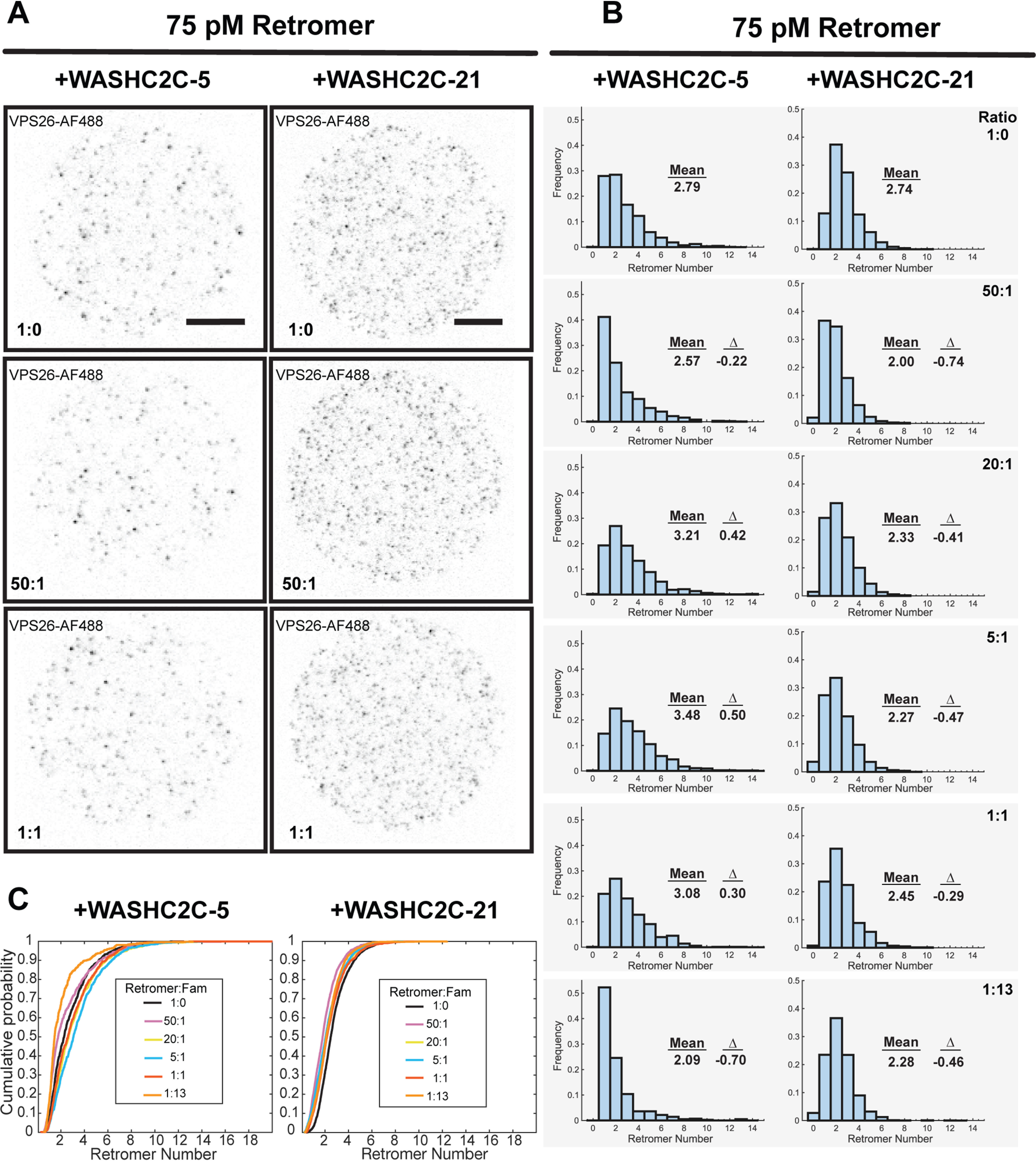
Figure 4.

WASHC2C does not influence Retromer oligomeric state on a supported bilayer. Single particle TIRFM analysis was used to measure retromer cluster size in the presence of varying amounts of WASHC2C proteins. His-tagged, Alexa Fluor 488–labeled retromer was attached to a SLB (solution concentration 75 pm) and the indicated Alexa Fluor 647–labeled WASHC2C protein was then added to the indicated retromer:WASHC2C ratio. After incubation, the SLB was washed and retromer was imaged. A, the left column of images shows retromer fluorescence after incubation with WASHC2C-5 (containing 5 LFa motifs) and the right column shows incubations with WASHC2C-21 (containing 21 LFa motifs). The number of retromer complexes in single puncta was calculated and the distribution was plotted. Retromer fluorescence is shown (gray values inverted). Scale bar: 10 μm. B, distributions of retromer oligomer size on the SLB after incubation with WASHC2C-5 or -21 at the indicated ratios. The mean for each distribution is indicated. In addition, the difference of the mean with the distribution obtained in the absence of WASHC2C fragments (1:0), Δ, is shown in each panel. The presence of WASHC2C shifted the mean by less than one retromer per cluster. Small shifts in the monomer-to-oligomer ratios are likely insignificant as well, because such differences are observed among technical or biological replicates (e.g. compare the first panels with 1:0, in the absence of WASHC2C fragments) C, distributions shown in B, were plotted as empirical cumulative distribution functions.
