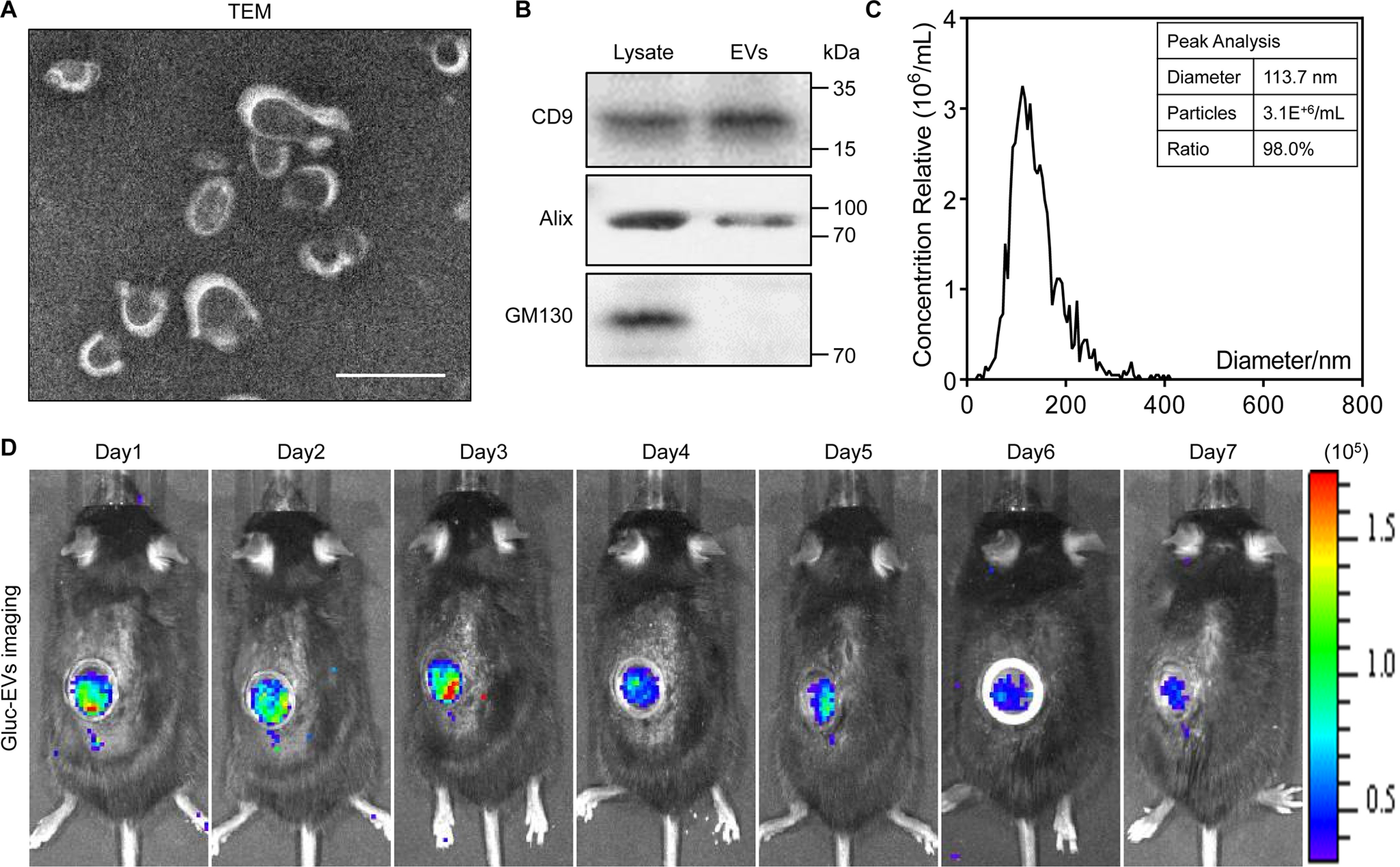
Figure 1.

Characteristics and bioluminescence imaging of EVs. A, transmission electron microscope (TEM) image of EVs. Scale bar, 100 nm. B, Western blot analysis confirmed the three categories of exosomal markers: CD9, Alix, and GM130. C, nanoparticle tracking analysis indicates the peak diameter of EVs is 113.7 nm. D, the biodistribution of EVs was traced in vivo by Gaussia luciferase (Gluc) imaging through the AIW.
