Figure 5.

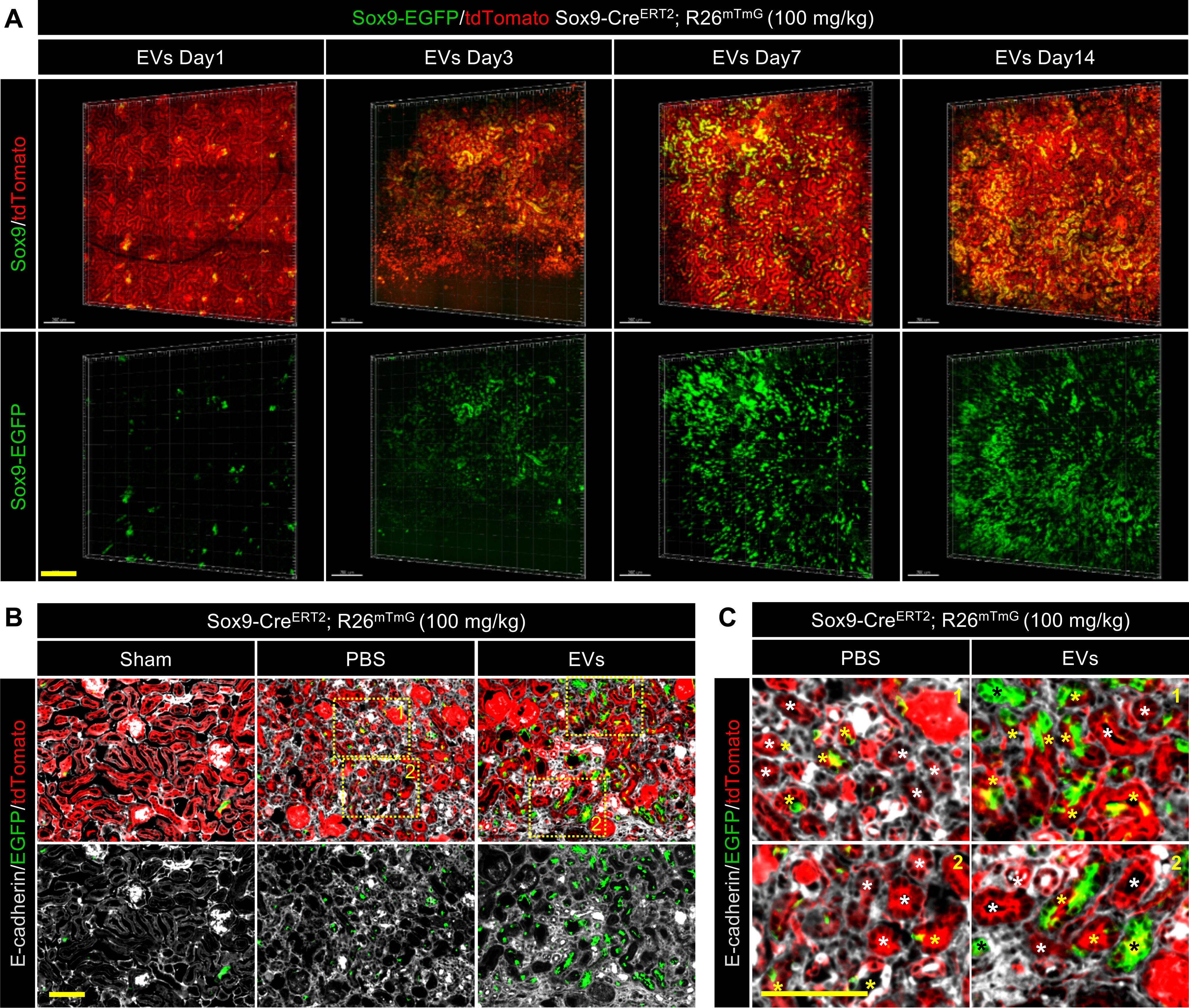
EVs promoted the formation of functional renal tubules by descendants of the Sox9+ cells. A, 3D reconstruction of the dynamic variation in the injured kidney treated with EVs by using two-photon living imaging. Scale bar, 200 μm. B and C, representative images (B) and local zoom images (C) for co-localization analysis of anti-E-cadherin immunostaining (gray) and Sox9-CreERT2–activated EGFP fluorescence in kidneys at 14 days post injury. White asterisks highlighted the tubules formed by descendants of Sox9− cells. Yellow asterisks highlighted the tubules formed by descendants of both Sox9+ cells and Sox9− cells. Black asterisks highlighted the tubules formed by descendants of only Sox9+ cells. Scale bar, 100 μm.
