FIGURE 2.
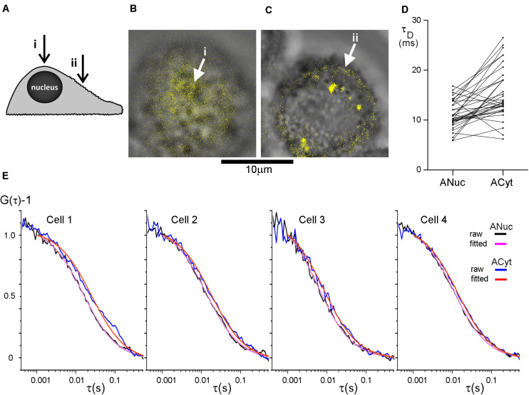
The diffusion in the plasma membrane appears to be faster on top of the nucleus than over the center of the cytoplasm. (A) The cells were subjected to FCS measurements at two different positions at their apical side plasma membranes; (i) on top of the nucleus and (ii) midway between the nucleus and the longest axes of the cell spread, above the cytoplasm center (indicated with arrows). HT29 cells stained with DiI imaged at (B) the top of the nucleus and (C) over the center of the cytoplasm with (i) and (ii) corresponding to panel (A). The DiI-signal in panels (B,C) was intensity thresholded and is displayed in false color. Scale bar 10 μm. (D) Comparison of the difference in τD from the plasma membrane on top of the nucleus and over the center of the cytoplasm. p = 5.0 × 10⁻⁶ for a two-tailed, paired t-test, and n = 36. Values from one cell were excluded from the graph, but not from the analysis, for clarity because its τD−values were considerably longer. (E) FCS curves from four different HT29 cells. On each cell FCS was measured above the nucleus (experimental curve in black, fitted curve in purple) and over the center of the cytoplasm (experimental curve in blue, fitted curve in red). Curves were fitted to a model assuming one diffusing species starting the fit at τ = 102 μs.

