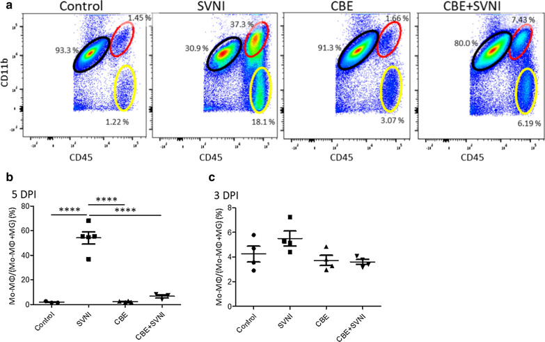
Fig. 5.
High levels of MΦ infiltration are observed in the brains of SVNI-infected mice. Data were obtained from C57BL/6 mice untreated (control) or treated with CBE (50 mg/kg per day) from 13 day of age, uninfected or infected with a lethal dose (30 pfu, 3LD50) of SVNI on 21 day of age. a Flow cytometry analysis of myeloid cells in the CNS of control, SVNI, CBE, and CBE + SVNI mice 5-DPI. Microglia express high levels of CD11b and intermediate levels of CD45 (indicated by the black ellipse), whereas monocyte-derived macrophage (Mo-MΦ) express high levels of both CD11b and CD45 (indicated by the red ellipse). Lymphocytes express high levels of CD45 and are negative for CD11b ((indicated by the yellow ellipse). n = 3–5 for each group. b, c The percentage of Mo-MΦ out of total myeloid lineage cells [Mo-MΦ + microglia (MG)] in the brain of control, SVNI, CBE, and CBE + SVNI mice 5- days (b) or 3-days (c) post infection. Pairs of proportions that resulted in both significant p values and large effect sizes (i.e., Cohen’s h > 0.8) were indicated according to their p values: *p < 0.05, ***p < 0.001, ****p < 0.0001

