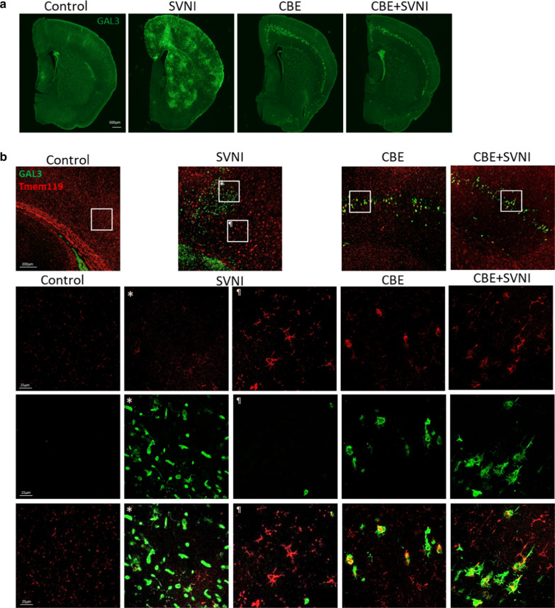
Fig. 6.
Distinct patterns of microglia activation in brains of SVNI-infected versus nGD mice. Data were obtained from C57BL/6 mice untreated (control) or treated with CBE (50 mg/kg per day) from 13 day of age, uninfected or infected with a lethal dose (30 pfu, 3LD50) of SVNI on 21 day of age. a Immunofluorescence of brains of control, SVNI, CBE, and CBE + SVNI mice using anti-GAL3 (green) antibody. GAL3 staining was evaluated using ImageJ. Control, SVNI, CBE, and CBE + SVNI contain ~ 308 ± 16, 1548 ± 644, 663 ± 46, and 713 ± 92 GAL3-positive cells, respectively. Results are representative of three biological replicates. b Double immunofluorescence of brains of control, SVNI, CBE, and CBE + SVNI mice using anti-GAL3 (green) and anti-Tmem119 (red) antibodies. ×2 (upper panel) and ×20 (lower panel) magnification images. Data were obtained 5-DPI. Results are representative of three biological replicates

