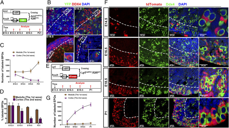
Fig. 5.
Lineage tracing of BPG cell replacement by cortical EPG cells. (A) Strategy for lineage-tracing bipotential cells. Mice containing Axin2CreERT2/+ mice and R26RYFP/YFP received tamoxifen (Tmx) at E10.5, and ovaries were analyzed at E12.5, E15.5, E19.5, and P21. (B) YFP-marked cells (green) are seen adjacent to germ cells (red) in the medulla and in the cortex until E19.5. Boxed regions correspond to Insets. (C) Quantitation of YFP-labeled BPG cells in the cortex and medulla at each time point shown in B. (D) Percentage of cyst/follicle-associated PG cells labeled in the medulla and cortex at each time. (E) Strategy for lineage tracing EPG cells. Mice containing Lgr5CreERT2/+ and R26RtdT/tdT reporter received Tmx at E13.5, and ovaries were analyzed at E14.5, E16.5, E18.5, and P1. (F) tdTomato-positive EPG cells (red) were sparse at E14.5 but associated with germ cells (green). Cortical tdTomato+ cells increased significantly at E16.5, E18.5, and P1, but very few entered the medullar region. Boxed regions correspond to Right Column. (G). Dashed lines in B and F show the boundary of the cortical and medullar regions.

