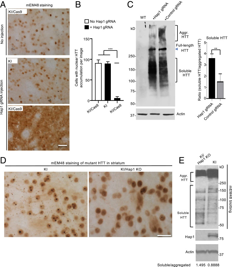
Fig. 5.
Loss of Hap1 in HD KI striatum reduces nuclear aggregated HTT and increases soluble mutant HTT. (A) mEM48 immunostaining showing that depletion of Hap1 by AAV-Hap1 gRNA injection reduced the accumulation of nuclear HTT and increased diffused mutant HTT in the striatum of a 6-mo-old KI/Cas9 mouse. The contralateral striatum region without injection in the same mouse and an age-matched KI mouse without Cas9 but injected with AAV-Hap1 gRNA served as controls. (B) Quantification of the number of cells with intense nuclear HTT accumulation. The data were obtained by counting 16 images (40X) from four animals per group. ***P < 0.001. (C) Western blotting (Left) shows the increased soluble mutant HTT and reduced HTT aggregates (Aggr. HTT) in the striatum of KI/Cas9 mice at 6 mo of age after AAV-Hap1 gRNA injection as compared with the control AAV-gRNA injection. (Right) The densitometric ratio of soluble HTT to aggregated (Agg.) HTT on Western blot (Left). The data are mean ± SEM (n = 3), **P < 0.01. (D) Tamoxifen-induced Hap1 depletion also reduced aggregated HTT and increased diffuse HTT distribution in the striatum in KI/Hap1 KO mice. KI/Hap1 KO mice were injected with tamoxifen at 3 mo of age and analyzed at 9 mo of age. (E) Western blotting also shows the reduced HTT aggregation (Aggr. HTT) and increased soluble HTT in the striatum when Hap1 was absent. The densitometric ratios of soluble HTT to aggregated HTT are shown beneath the Western blots. (Scale bars, 20 μm [A]; 15 μm [D].)

