Fig. 1.
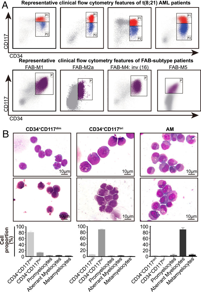
Clinical immunophenotypic characteristics and morphological features of distinct leukemic cell populations in t(8;21) AML patients. (A) Representative clinical flow cytometry data showing the distribution of CD34+ myeloblasts by antigen CD34 and CD117 in four t(8;21) AML patients at diagnosis (Upper) and four AML patients with other FAB subtypes (except for M3) at diagnosis (Lower). (B, Upper) Representative Wright Giemsa-stained cytospin preparations of isolated CD34+CD117dim, CD34+CD117bri, and AM cell populations from the BM of t(8;21) AML patients. The AM population was isolated using the marker combination of CD34−CD117−HLA-DR−CD15+CD11b−. (B, Lower) Differential counts of the isolated cell populations (mean ± SD; n = 4), including CD34+CD117dim, CD34+CD117bri, and AM populations.

