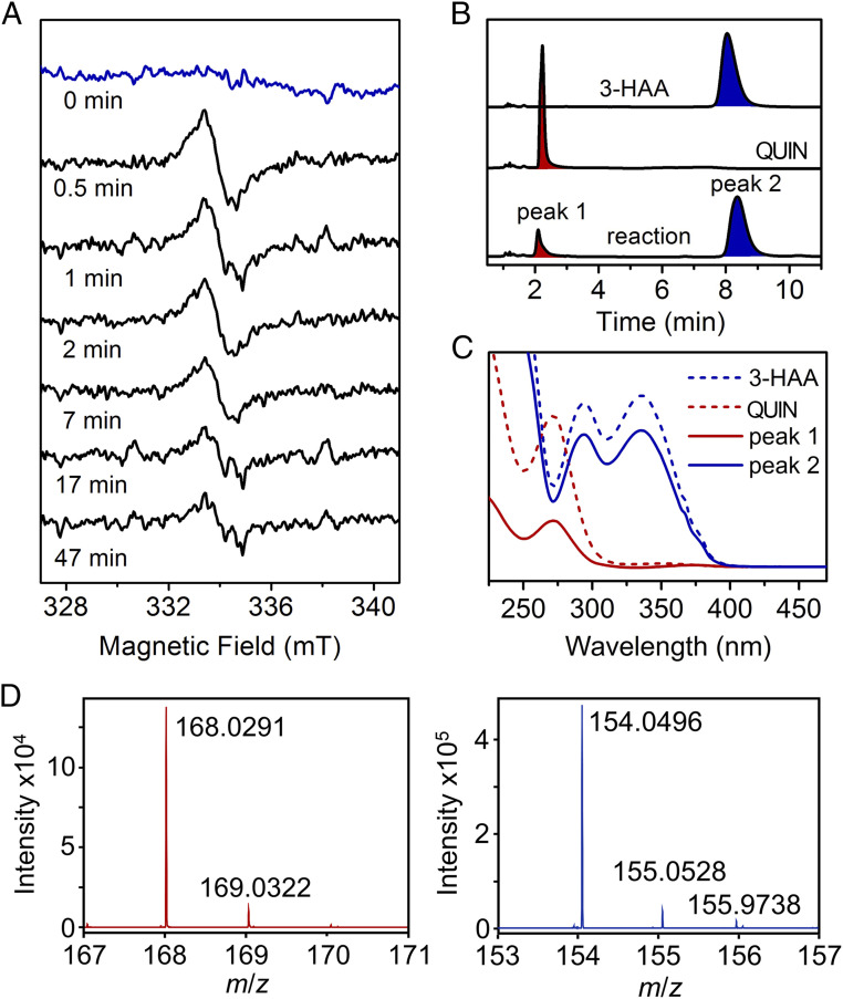
Fig. 2.
Single-crystal continuous-wave EPR spectroscopy of the HAO reaction with a crystal slurry reveals a substrate-based radical intermediate. (A) Time-lapse in crystallo reaction at room temperature with samples analyzed by X-band EPR spectroscopy at 70 K. (B) HPLC–UV-vis analysis of the single-crystal reaction mixture. Elution profiles are shown with the absorbance at 280 nm. (C) UV-vis spectra of the HPLC fractions eluted with solvent containing 2% acetonitrile and 0.1% formic acid. (D) High-resolution mass spectrometry of the HPLC fractions. Peak 1 (blue) was identified as 3-HAA, and peak 2 (red) was QUIN.

