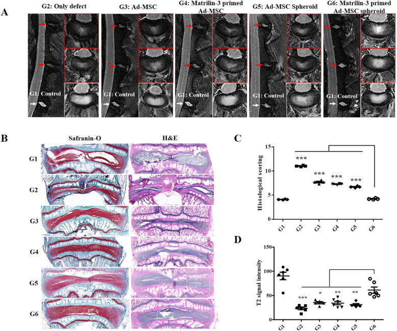
Fig. 5.
T2-weighted MRI and histological analysis for assessing disc regeneration in a rabbit model. a Representative T2-weighted MRI scans of the sham, single Ad-MSC (ASC), matrilin-3 (MATN3) + single ASC, ASC spheroid, and MATN3 + ASC spheroid groups. b Disc regeneration was assessed by Masson’s trichrome staining. The results revealed that disc regeneration was the highest in the matrilin-3-primed ASC spheroid group. c The histological score for disc regeneration was the highest in the matrilin-3-primed ASC spheroid group. d The T2 signal intensity was the highest in the matrilin-3-primed ASC spheroid group, suggesting that the matrilin-3-primed ASC spheroid was the most effective for IVD regeneration (n = 7). G1, control; G2, only defect; G3, single Ad-MSC; G4, matrilin-3-primed single Ad-MSC; G5, Ad-MSC spheroid; G6, matrilin-3-primed Ad-MSC spheroid. ***p < 0.001, **p < 0.01, *p < 0.05

