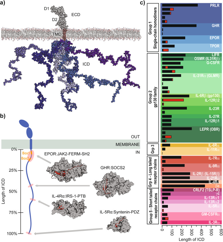
Fig. 1.
The C1CR family - structures and ICD isoforms. a Structural model of the full-length human PRLR [35] in the membrane, with the ECD in light grey, the TMD in pink and a total of six ICD chains shown in blue shades to represent its disordered conformational ensemble. b Left: A representative sketch of a prototypical C1CR (blue) in a membrane (green) having a long ICD with its length given in %. Box1 and Box2 are highlighted in orange. The red shades highlight the approximate extent and position of small ICD fragments of different C1CR-ICDs having their structure solved in complex with signaling proteins. Right: Three-dimensional structures of signaling proteins (grey surfaces) in complex with C1CR-ICD fragments (red cartoon and sticks), being the FERM-SH2 domain of JAK2 in complex with EPOR-ICD279–334 (pdb code 6E2Q) (top), SOCS2 in complex with GHR-ICD591–603 (pdb code 6I5J, 6I5N) (middle top), the PTB domain of IRS-1 in complex with IL-4Rα490–500 (pdb code 1IRS) (middle bottom) and the PDZ domain from syntenin in complex with IL-5Rα417–420 (pdb code 1OBZ) (bottom). The grey arrows point to the approximate positions of the ICD fragments (red shades) on the representative ICD sketch. c The 29 receptors of the C1CR family with an ICD and their isoforms. The length of the ICDs are to scale and red indicates the length of unique sequences differing from the long-form. The receptors are divided into 5 groups according to [33].

