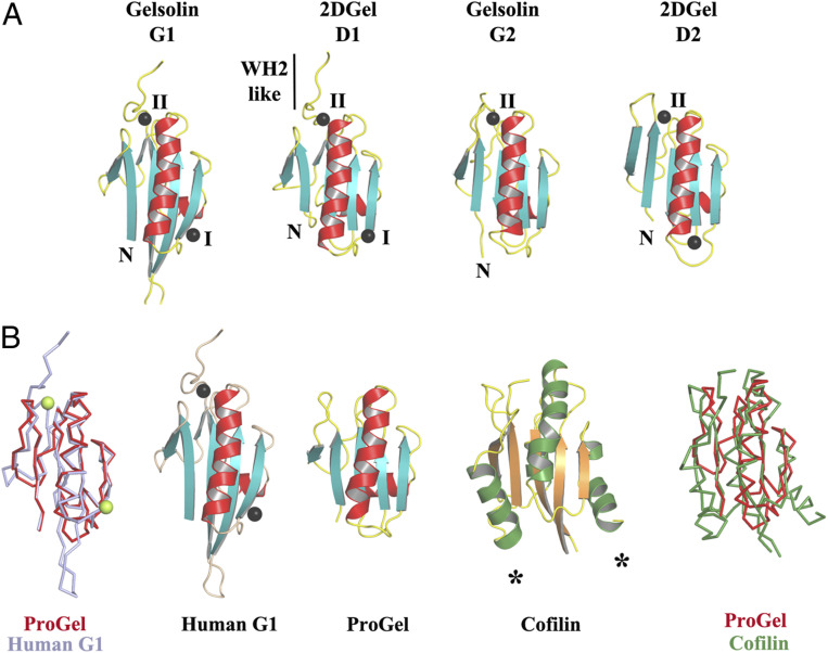
Fig. 5.
Structural homology to gelsolin and cofilin. (A) The three conserved calcium-binding sites in 2DGel and 2DGel3 are shared with the first two domains of human gelsolin. (B) Structural comparisons and superimpositions of ProGel with human gelsolin G1 (PDB ID code 3FFN) and cofilin (PDB ID code 4KEE). Calcium ions are shown as lime or black spheres. Asterisks indicate additional helices in the cofilin fold relative to ProGel.

