Figure 1.
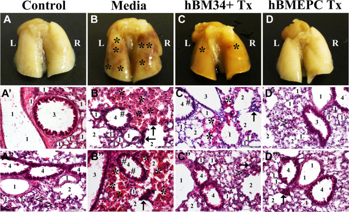
Characteristic of the lungs in G93A SOD1 mice at the late disease stage. Gross view of the lungs with dorsal side up showed abundant microhemorrhages (mh) in media-treated mice (B) versus controls (A). At 4 wk post-treatment, a substantial decrease of mh was noted after hBM34+ cell treatment (C) whereas no mh were found in ALS mice receiving hBM-EPCs (D). The H&E staining in left lung lobe revealed typical appearance of all lung compartments, including cellular components in control mice (A′, A″). In contrast, diffuse eosinophilic infiltration around bronchioles and bronchiolar epithelium damage were seen in media-treated mice (B′, B″). Also, ruptured capillaries near alveoli or alveolar sacs were noted. Mice treated with hBM34+ cells demonstrated some areas of eosinophilic permeation, mild damage of bronchiolar epithelium, and a few burst capillaries (C′, C″). Near normal presence of airway components was observed in ALS mice receiving hBM-EPCs (D′, D″). Only scarce ruptured capillaries around alveolar sacs were found (D″). Scale bar in A′–D″ is 100 µm. L: left lobe; R: right lobe; 1: alveolus; 2: alveolar sac; 3: bronchus; 4: bronchiole; <: typical capillary; ←: ruptured capillary; *: microhemorrhage; #: damaged bronchiolar epithelium; Tx: cell transplant; ALS: amyotrophic lateral sclerosis; hBM34+: human bone marrow-derived CD34+ cells; hBM-EPCs: human bone marrow endothelial progenitor cells; H&E: hematoxylin & eosin.

