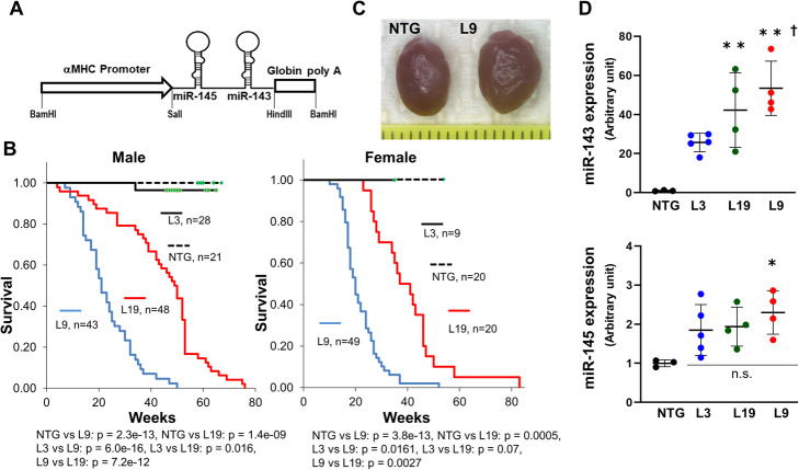
Fig. 1.
Establishment of αMHC/miR-143/145TG mice. a Structure of the injected fragment. An approximately 6.7-kb BamHI fragment containing the pri-miR-145 and pri-miR-143 genes was used. b Kaplan-Meier survival analysis of αMHC/miR-143/145TG mice. Data were analyzed using a log-rank test followed by a post hoc Holm test. Green squares = censored. c Hearts from representative 5-month old male NTG and L9 mice. The scale bar is 1 mm. d Quantitative RT-PCR analysis of miR-143 and miR-145 in the hearts of 3-month old male αMHC/miR-143/145TG mice. The results are presented as the means ± SD with scattered blots. Significance was assessed with one-way ANOVA followed by a post hoc Tukey test (n = 3 ~ 5. *p < 0.05 vs. NTG; **p < 0.01 vs. NTG; †p < 0.05 vs. L3). Similar results were obtained in at least two independent experiments

