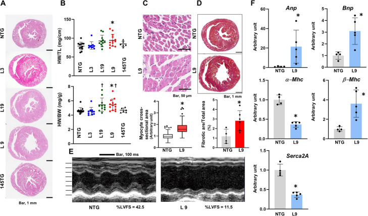
Fig. 2.
Characterization of the hearts in αMHC/miR-143/145TG and αMHC/miR-145TG mice. a Macroscopic histological analysis of hematoxylin & eosin-stained hearts of 4-month old male αMHC/miR-143/145TG and αMHC/miR-145 TG mice. b Heart weight corrected for tibia length (upper panel) or body weight (lower panel) of male αMHC/miR-143/145TG and αMHC/miR-145TG mice. The results are presented as the means ± SD with scattered blots. Significance was assessed with one-way ANOVA followed by a post hoc Tukey test (n = 6 ~ 13; *p < 0.05 vs. NTG, †p < 0.05 vs. L3). c Microscopic histological analysis of hematoxylin & eosin-stained hearts of 4-month old male L9 mice. The lower panel shows the quantitative analysis of the myocyte cross-sectional area. We analyzed 316 cardiomyocytes of four L9 mice at 4 months of age and 292 cardiomyocytes of three NTG mice. Data are presented as box and whisker plots with the Tukey method and unpaired t-test applied (*p < 0.05 vs. NTG). d Macroscopic histological analysis of Masson-trichrome-stained hearts of 4-month old male L9 mice. The lower panel shows the quantitative analysis for the fibrotic area. The results are presented as the means ± SD with the unpaired t-test applied to determine significance (n = 4 ~ 6; *p < 0.05 vs. NTG). e Representative M-mode echocardiography of a 3-month old male L9 mouse. The interval of each scale bar of the Y-axis is 1 mm. %LVFS: percentage left ventricular fractional shortening. f Quantitative RT-PCR analysis of molecules correlating with cardiac remolding of 3-month old male L9 mice. The results are presented as the means ± SD with the unpaired t-test applied to determine significance (n = 4 ~ 5; *P < 0.05 vs. NTG). Similar results were obtained in at least two independent experiments

