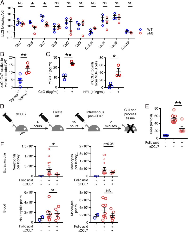
FIGURE 4.
Provision of CCL7 from B cells attracts inflammatory myeloid cells into the kidneys during AKI. (A) qPCR from whole kidney lysate from WT and μMt mice (blue and red bars, respectively) 15 h following FA-AKI showing CCL5 and CCL7 significantly reduced in μMt mice (n = 6 male mice in total); a parametric unpaired t test was used. (B) Ccl7 transcript levels in kidney lysate 15 h following FA-AKI in WT and Siglecg−/− mice (n = 9 male mice in total); a parametric unpaired t test was used. (C) Supernatant from negatively isolated WT B cells were measured for CCL7 by ELISA following stimulation with CpG (left). Supernatants from MD4 B cells were measured for CCL7 following BCR ligand stimulation with HEL (right, [n = 3 male mice in each experiment]; 5 × 10⁵ B cells stimulated for 48 h; a parametric unpaired t test was used.) (D). Schematic of experiment inhibiting CCL7 during AKI. (E) Serum urea following with CCL7 inhibition (n = 16 male mice in total); two identical experiments were combined; and a parametric unpaired t test was used. (F) Extravascular renal and blood neutrophils and monocytes following CCL7 inhibition. *p < 0.05, **p < 0.01.

