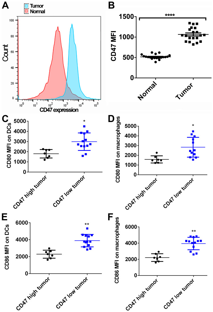
Figure 1.
Expression of CD47 and activation of antigen-presenting cells in PDAC samples. (A) CD47 expression in normal pancreatic and paired tumor tissues was measured by fluorescence-activated cell sorting analysis (sample size=20). Tumor tissues exhibited higher CD47 expression as compared with that in normal tissues. (B) Quantified data showing that the MFI of CD47 in tumor tissues was higher than that in normal tissues. (C) DCs and (D) macrophages exhibited higher CD80 expression in human PDAC samples with low CD47 expression. (E) DCs and (F) macrophages exhibited higher CD86 expression in human PDAC samples with low CD47 expression. *P<0.05, **P<0.01 and ****P<0.0001. CD47, cluster of differentiation 47; PDAC, pancreatic ductal adenocarcinoma; MFI, mean fluorescence intensity; DCs, dendritic cells.

