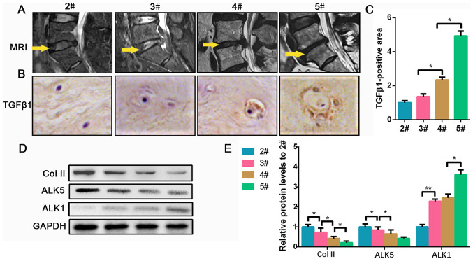
Figure 1.
TGF-β1 expression is increased in degenerated discs. (A) Representative MRI of the patients from Pfirrmann grade 2 to 5. The yellow arrow indicates the surgical segment. (B) Representative images of immunohistochemical targets TGF-β1 (magnification, x400) and (C) quantification analysis. (D) Protein expression of Col II, ALK5 and ALK1 was determined by western blotting and (E) semi-quantified. Data are presented as the mean ± SD of three independent experiments. *P<0.05, **P<0.01. TGF-β1, transforming growth factor β1; Col II, collagen II; MRI, magnetic resonance imaging; ALK, ALK tyrosine kinase receptor.

