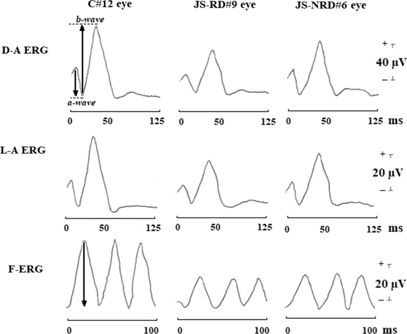
Fig. 1.
Examples of dark-adapted electroretinogram (D-A ERG), light-adapted ERG (L-A ERG), and 30-Hz flicker ERG (F-ERG) responses assessed in one control subject (C#12 eye), in one representative patient with Joubert syndrome with retinal dystrophy (JS-RD#9 eye), and in one representative patient with Joubert syndrome without retinal dystrophy (JS-NRD#6 eye). With respect to control eye, both JS-RD and JS-NRD eyes showed light-adapted ERG and dark-adapted ERG with a delay in a- and b-wave implicit times (dashed lines) and with a reduction in a- and b-wave peak-to-peak amplitudes (arrows) and a reduction of F-ERG peak-to-peak amplitudes (arrows). ms milliseconds, µV microvolt

