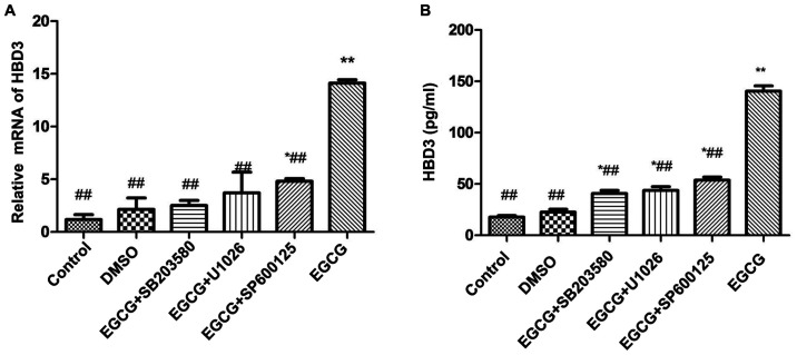
Figure 4.
Effects of EGCG on the expression of HBD3 in BEAS-2B cells. (A) Real-time RT-PCR assay for the upregulated expression of HBD3 mRNA treated with inhibitors for intracellular pathways. The cells were treated with inhibitors for intracellular pathways in a series of experiments. BEAS-2B cells were pre-incubated with SB203580 (an inhibitor of p38 MAPK), U1026 (an inhibitor of ERK), and SP600125 (an inhibitor of JNK) for 1 h and then treated with 50 µg/ml EGCG for 12 h. SB203580 (an inhibitor of p38 MAPK), U1026 (an inhibitor of ERK), and SP600125 (an inhibitor of JNK) completely inhibited the upregulated expression of HBD3 by EGCG (*P<0.05, **P<0.01, compared with the control group; #P<0.05, ##P<0.01 compared with the EGCG group). The data are representative of three independent experiments. (B) Competitive ELISA for the quantification of HBD3 secretion by cultured BEAS-2B cells after EGCG and inhibitor treatments. The cell were preincubated with SB203580, U1026, and SP600125 for 1 h and then treated with 50 μg/ml EGCG for 12 h. Serial dilutions of synthetic mature HBD3 peptide were used to generate a standard curve, and the HBD3 peptide concentration of each experimental group was considered. In accordance with the RT-PCR result, SB203580, U1026, and SP600125 completely inhibited the secretion of HBD3 by EGCG (*P<0.05, **P<0.01, compared with the control group; ##P<0.01, compared with the EGCG group). The data are representative of three independent experiments. EGCG, Epigallocatechin gallate.

