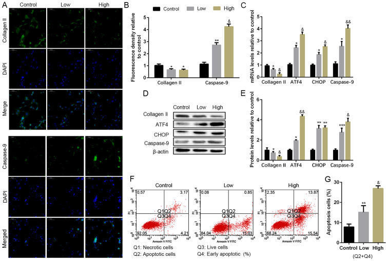
Figure 2.
Apoptosis levels in H2O2 induce NP cells in vitro. NP cells of G2 tissues were cultured with a low or high concentration of H2O2 (50 or 100 µM) for 24 h. (A and B) The protein expression level of collagen II and caspase-9 were determined by (A) IF (magnification, x400) and (B) quantification analysis. (C) The mRNA expression levels of collagen II, ATF4, CHOP, and caspase-9 were assayed by RT-PCR. (D and E) The protein expression levels of collagen II, ATF4, CHOP and caspase-9 were determined by (D) WB and (E) quantification analysis. (F and G) The ratio of apoptotic cells was analyzed by (F) flow cytometry and (G) quantification analysis. The values are mean ± SD of three independent experiments (n=3). (*P<0.05, **P<0.01, ***P<0.001, compared with control; &P<0.05, &&P<0.01, compared with 50 µM H2O2). ATF4, activating transcription factor 4; CHOP, C/EBP homologous protein; WB, western blot; NP, nucleus pulposus; RT-PCR, reverse transcription-polymerase chain reaction; IF, immunofluorescence.

