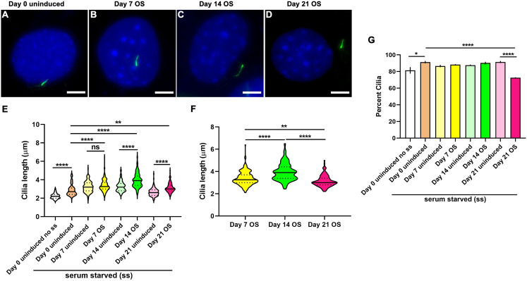
Figure 5. Osteogenic differentiation in MC3T3-E1 preosteoblast cells caused a distinct pattern of cilia elongation and reduction in frequencies.
(A–D) Representative images of MC3T3-E1 primary cilium at day 0 uninduced and days 7, 14 and 21 after CH induction. Primary cilia were labeled with acetylated α tubulin (green), while nuclei were stained with DAPI (blue). Scale bar: five μm. All OS treated and untreated cells were serum starved (ss) except one set of uninduced cells at day 0 (day 0 uninduced no ss). (E) Serum starvation caused cilia length increase at day 0; at day 7 after osteogenesis cilia were longer than day 0 uninduced; at 14 and 21 days following OS differentiation the cilia are significantly longer than both day 0 and their corresponding day matched controls. n = 101–178 (**p < 0.01, ****p < 0.0001, One-way ANOVA followed by Tukey’s post hoc test). (F) Among OS media induced cells cilia were longest at 14 days of OS differentiation (**p < 0.01, ****p < 0.0001, Two-way ANOVA followed by Tukey’s post hoc test). (G) Starvation appeared to increase primary cilia prevalence in all induced and uninduced monolayers. However, cilia frequencies were significantly reduced only in 21 days OS differentiated cells, n = 326–710 (*p < 0.05, ****p < 0.0001; One way ANOVA followed by Tukey’s post hoc test).

