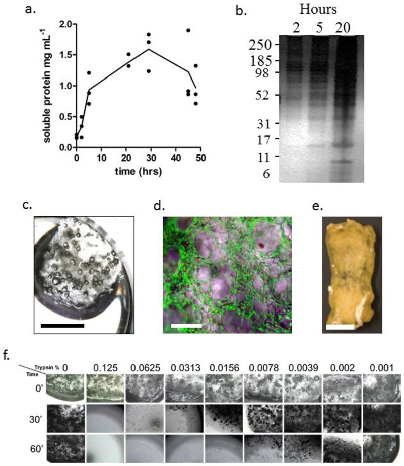
Figure 3: Solubilization and coating of OECM:
Panel a: Plot of solubilization of OECM over time in acetic acid. Panel b: Silver stained SDS-polyacrylamide electrophoresis of solubilized OECM. Numbers of left represent approximate molecular mass in kilodaltons. Panel c: GF fragment in 12-well culture dish for purposes of MSC culture and OECM coating. Panel d: Composite phase-fluorescence micrograph of live-dead stained fragment of GF illustrating attached live MSCs (green) and occasional dead MSC nuclei (red). Panel e: Bioconditioned GF construct for the purposes of a bone repair assay in mice. OECM is incorporated throughout the thickness of the bioconditioned construct, and has the handling properties of decellularized bone (adapted with permission from ref.48). Panel f: Dose response experiment demonstrating the sensitivity of unmodified GF to trypsin. Note that the construct in Panel e was incubated in 0.125 (v/v) trypsin for 15 hr. For panel c,e: bar = 5 mm. For panel d: bar = 100 μm.

