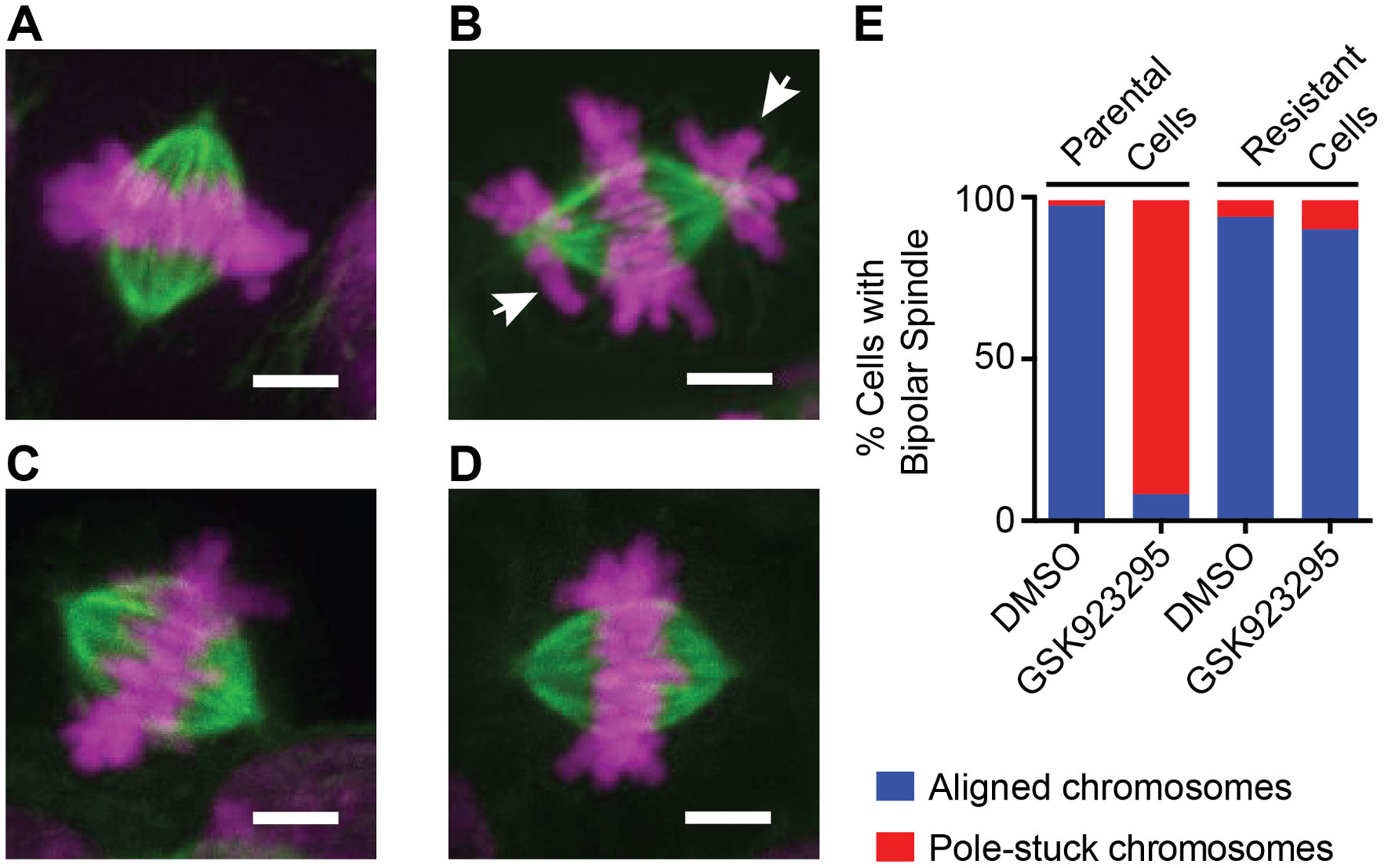
Figure 3. Immunofluorescence analysis of parental and GSK923295-resistant diploid cells.

(A-D) Representative immunofluorescence images of mitotic spindles in parental and GSK923295-resistant HCT116 cells. Maximum intensity projections of DNA (magenta) and tubulin (green) are shown. Parental cells treated with vehicle control (A) or GSK923295 (B), and GSK923295-resistant cells (clone 2) treated with vehicle control (C) or GSK923295 (D) are shown. Pole-stuck chromosomes are indicated with white arrows. Scale bars, 4 μm.
(E) Bipolar spindles were counted and manually classified as aligned or containing pole-stuck chromosome(s). Spindles in parental cells treated with DMSO: aligned: 98%, pole-stuck: 2%; parental cells treated with GSK923295: aligned: 8%, pole-stuck: 92%; spindles in resistant cells (clones 1 and 2) treated with DMSO: aligned, 95%; pole-stuck, 5%; GSK923295: aligned, 91%; pole-stuck, 9%. See also Figure S3.
