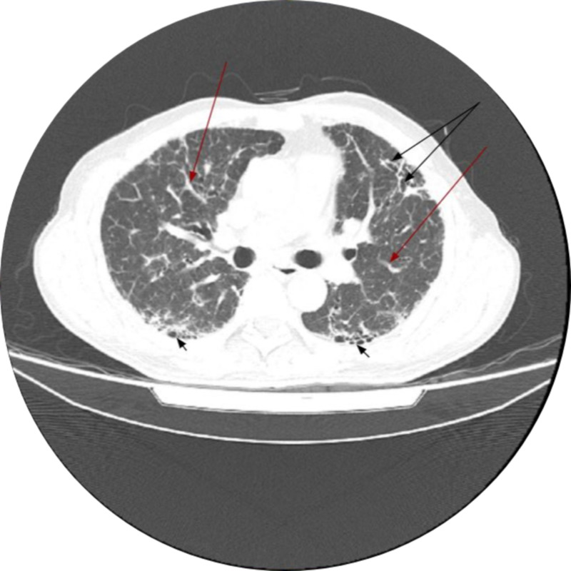
Figure 1. High-resolution computed tomographic features of usual interstitial pneumonia.
Diffuse interlobar interstitial thickening is noted at bilateral aerated lungs (red arrows) along with honeycombing more prevalent at lung bases (short black arrows) and mild traction bronchiectasis at left upper lobe (longer black arrows at the top)

