Figure 3. a–d.

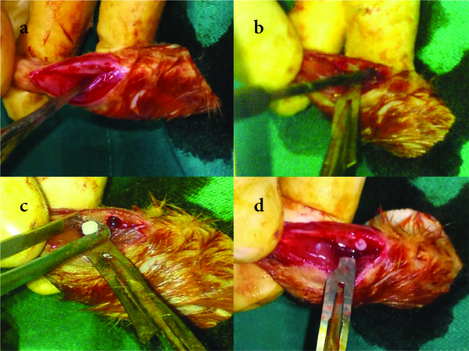
Intraoperative photos representing (a) the dissected medial tibial cortex, (b) the drilling of the tibia, (c) the implant placement, and (d) the tibial fracture model with an implant in place before skin closure

Intraoperative photos representing (a) the dissected medial tibial cortex, (b) the drilling of the tibia, (c) the implant placement, and (d) the tibial fracture model with an implant in place before skin closure